SRT-100
2020年12月美国Sensus Healthcare正式授权Ekpac Healthcare(香港维昌医疗)为SRT-100中国大陆及香港地区的独家代理
环钻切除术在治疗胸部多发性瘢痕疙瘩中的临床应用
刘龙灿 鲜华 敖贤 许丹 安娟 王春梅
本文来源:《中华整形外科杂志》2023年12月 第39卷 第12期
DOI:10. 3760 / cma.j.cn114453-20230301-00044
作者单位:南方医科大学皮肤病医院整形美容外科, 广州510091
通信作者:王春梅,Email:1944134044@qq.com
引用本文
刘龙灿,鲜华,敖贤,等. 环钻切除术在治疗胸部多发性瘢痕疙瘩中的临床应用[J]. 中华整形外科杂志,2023,39(12):1277-1283. DOI:10.3760/cma.j.cn114453-20230301-00044
【摘要】
目的 探讨环钻切除术加浅层X线照射联合局部药物注射治疗胸部多发性瘢痕疙瘩的可行性和临床效果。
方法 选取2020年3月至2021年6月于南方医科大学皮肤病医院治疗的胸部多发性瘢痕疙瘩患者,采用随机数字表法分成观察组和对照组。观察组先进行环钻切除术,术后24 h内照射1次浅层X线,之后每周照射1次,共照射4次,放疗结束1周后予局部药物注射治疗(1 ml醋酸曲安奈德注射液+0.6 ml氟尿嘧啶注射液+3.4 ml 2%盐酸利多卡因注射液制备成5 ml混合液),每月1次,共4次,单次注射终点反应为瘢痕疙瘩变白;对照组不接受环钻切除术,其他治疗与观察组相同。于治疗前和治疗后3、6、12个月进行温哥华瘢痕量表(VSS)评分和治疗有效性评价(治愈、显效、好转、无效),记录治疗前后瘢痕疙瘩总体积、局部注射药物总量和不良反应情况。采用SPSS 26.0软件进行数据分析,计量资料数据用x±s表示,2组间比较采用独立样本 t检验,同组治疗前后比较采用重复测量数据的方差分析;计数资料用百分数表示,组间比较采用 χ 2检验。P<0.05为差异有统计学意义。
结果 共纳入58例患者,每组各29例,其中男性36例,女性22例;年龄18~59岁,平均29岁;每例患者胸部瘢痕疙瘩数量为5~12个。治疗前,观察组与对照组VSS评分差异无统计学意义[(13.21±1.24)分vs.(12.90±1.21)分, t=0.97, P=0.337]。治疗结束后随访3、6、12个月,观察组VSS评分分别为(4.21±1.26)、(4.34±1.40)、(4.55±1.33)分,对照组VSS评分分别为(5.66±1.32)、(6.07±1.44)、(6.62±1.40)分,2组间比较差异有统计学意义( t=-4.27、-4.63、-5.78, P均<0.001);组内比较显示,观察组患者治疗后3、6、12个月VSS评分差异无统计学意义( F=2.50, P=0.111),对照组患者治疗后3、6、12个月VSS评分呈逐渐升高趋势,差异有统计学意义( F=30.75, P<0.001)。观察组显效22例,好转7例,显效率为75.86%(22/29);对照组显效6例,好转23例,显效率为20.69%(6/29),2组显效率差异有统计学意义( χ 2=17.68, P<0.001)。治疗前观察组与对照组瘢痕疙瘩总体积差异无统计学意义[(7.76±1.71) cm 3 vs.(8.27±1.26) cm 3, t=-1.28, P=0.207];治疗后随访12个月,观察组瘢痕疙瘩总体积[(2.57±0.59) cm 3]明显小于对照组[(5.51±1.39) cm 3],差异有统计学意义( t=-10.47, P<0.001)。观察组局部注射药物总量[(6.45±1.25) ml]明显少于对照组[(11.00±1.73) ml],差异有统计学意义( t=-11.48, P<0.001)。2组患者均在进行浅层X线照射后3~5 d出现照射区域一过性色素沉着,6个月内自然消退。除此之外,观察组出现5例不良反应,分别为部分创面延迟愈合2例,毛细血管扩张2例,月经周期异常1例;对照组出现13例不良反应,包括毛细血管扩张7例,月经周期异常2例,皮肤萎缩凹陷4例。观察组不良反应发生率明显低于对照组,差异有统计学意义[17.24%(5/29) vs. 44.83%(13/29), χ 2=5.16, P<0.001]。
结论 环钻切除术加浅层X线照射联合局部药物注射治疗胸部多发性瘢痕疙瘩,具有操作简便、治疗效果好、不良反应少的优点。
【关键词】瘢痕疙瘩;环钻;浅层X线;局部注射
Clinical application of punch excision in the treatment of chest multiple keloids
Liu Longcan, Xian Hua, Ao Xian, Xu Dan, An Juan, Wang Chunmei
Department of Plastic & Cosmetic Surgery, Dermatology Hospital of Southern Medical University, Guangzhou 510091, China
Corresponding author: Wang Chunmei, Email: 1944134044@qq.com
【Abstract】
Objective To explore the feasibility and clinical outcomes of punch excision combined with superficial X-ray and intralesional drug injection in the treatment of chest multiple keloids.
Methods Patients with chest multiple keloids in Dermatology Hospital of Southern Medical University from March 2020 to June 2021 were selected and randomly divided into observation group and control group according to random number table. In the observation group, punch excision was performed first, superficial X-ray was irradiated once within 24 h after surgery, and then once a week, for a total of 4 sessions. One week after radiotherapy, intralesional injection was performed with 5 ml mixture of 1 ml triamcinolone acetate injection, 0.6 ml fluorouracil injection and 3.4 ml 2% lidocaine hydrochloride injection, once a month, for a total of 4 times. Single injection endpoint reaction was keloid whiteness. The control group did not receive punch excision, and other treatments were the same as those in the observation group. Vancouver scar scale (VSS) score and efficacy evaluation (cure, excellent, improvement, ineffective) were performed before treatment and 3, 6, 12 months after treatment. Total keloids volume, intralesional injection volume and adverse reactions before and after treatment were recorded. SPSS 26.0 software was used for data analysis, and the measurement data were expressed as Mean±SD. Independent sample t-test was used for comparison between two groups, and repeated measurement data analysis of variance was used for comparison before and after treatment in the same group. Statistical data were expressed as % and χ 2 test was used for comparison between groups. P<0.05 was considered statistically significant.
Results A total of 58 patients were included, 29 in each group. There were 36 males and 22 females. The age ranged from 18 to 59 years old, with an average age of 29. The number of chest keloids in each patient was 5-12. Before treatment, the VSS scores of observation group and control group was 13.21±1.24 and 12.90±1.21 respectively. There was no significant difference in VSS scores between the two groups before treatment ( t=0.97, P=0.337). After 3, 6 and 12 months of follow-up, the VSS scores of the observation group were 4.21±1.26, 4.34±1.40 and 4.55±1.33 respectively, while those of the control group were 5.66±1.32, 6.07±1.44 and 6.62±1.40 respectively. The differences between the two groups were statistically significant ( t=-4.27, -4.63, -5.78, all P<0.001). Intra-group comparison showed that there were no statistically significant differences in VSS scores at 3, 6 and 12 months after treatment in the observation group ( F=2.50, P=0.111), while VSS scores at 3, 6 and 12 months after treatment in the control group showed a gradually increasing trend, with statistically significant difference ( F=30.75, P<0.001). In the observation group, 22 cases showed excellent effect, 7 cases improved, and the excellent rate was 75.86%(22/29). In the control group, there were 6 cases of excellent effect and 23 cases of improvement, the excellent rate was 20.69%(6/29), and the difference between the two groups was statistically significant ( χ 2=17.68, P<0.001). Before treatment, the total keloids volume of the observation group was (7.76±1.71) cm 3, which was (8.27±1.26) cm 3 of the control group, and there was no significant difference between the two groups before treatment ( t=-1.28, P=0.207). In the follow-up of 12 months after treatment, the total keloids volume of the observation group[(2.57±0.59) cm 3] was significantly smaller than that of the control group[(5.51±1.39) cm 3], and the difference was statistically significant ( t=-10.47, P<0.001). The total amount of intralesional injection in the observation group[(6.45±1.25) ml] was less than that in the control group[(11.00±1.73) ml], and the difference was statistically significant ( t=-11.48, P<0.001). Transient hyperpigmentation appeared in the irradiation area 3-5 days after superficial X-ray, which subsided naturally within 6 months in both groups. In addition, there were 5 adverse reactions in the observation group, including delayed healing (2 cases), telangiectasia (2 cases) and abnormal menstrual cycle (1 case). There were 13 cases of adverse reactions in the control group, including telangiectasia (7 cases), abnormal menstrual cycle (2 cases), and atrophic and sunken skin (4 cases). The incidence of adverse reactions in the observation group was significantly lower than that in the control group, and the difference was statistically significant [17.24%(5/29) vs. 44.83%(13/29), χ 2=5.16, P<0.001].
Conclusion Punch excision combined with superficial X-ray and intralesional drug injection for the treatment of chest multiple keloids has the advantages of simple operation, good therapeutic effect and less adverse reactions.
【Key words】Keloid; Punch; Superficial X-ray; Local injection
Disclosure of Conflicts of Interest: The authors have no financial interest to declare in relation to the content of this article.
Ethical Approval: Ethical approval was given by the Medical Ethics Committee of Dermatology Hospital of Southern Medical University (2021003).
瘢痕疙瘩是继发于皮肤外伤或自发形成的过度生长的病理性瘢痕,其治疗手段多种多样,但单一治疗的复发率较高,成为困扰临床医师的难题 [ 1 ]。多发性瘢痕疙瘩是指同一例患者多个解剖部位出现的多个瘢痕疙瘩,多继发于毛囊炎、痤疮等皮肤炎症 [ 2 ],治疗更具挑战性。手术联合浅层X线照射治疗已被证实是瘢痕疙瘩的一线治疗方案 [ 3 , 4 ],但当多个瘢痕疙瘩紧密相连、数量较多时,很难对每个瘢痕疙瘩进行针对性手术,且当患者出现大面积瘢痕疙瘩(超过10 cm×10 cm)时,也不适宜采用传统的手术方式。因此,多发性瘢痕疙瘩的治疗策略为软化、淡化瘢痕和缓解痛痒症状,局部皮损内注射是治疗多发性瘢痕疙瘩的主要手段 [ 5 ]。我们团队的前期研究表明,对于多发性瘢痕疙瘩,局部药物注射前进行浅层X线照射治疗的效果优于单纯注射 [ 6 ]。但是在不进行手术减容的前提下,应用局部药物注射联合浅层X线照射治疗多发性瘢痕疙瘩,需要的注射药物量大、疗程长,这迫使我们思考更佳的治疗方法。环钻切除术是利用环钻器钻孔切除皮肤组织的一种技术,切除后的创面呈圆形,具有操作简便、损伤小的特点。我们应用环钻切除术加浅层X线照射联合局部药物注射治疗胸部多发性瘢痕疙瘩,并探讨其可行性和临床疗效,以期为临床实践提供新的安全有效的治疗方案。
资料与方法
一、病例选择
选取2020年3月至2021年6月于南方医科大学皮肤病医院进行治疗的胸部多发性瘢痕疙瘩患者,采用随机数字表法分成2组,观察组采用环钻加浅层X线照射联合局部药物注射治疗,对照组采用浅层X线照射联合局部药物注射治疗。
纳入标准:(1)继发于皮肤感染的胸部多发性瘢痕疙瘩,瘢痕疙瘩数量≥5个;(2)温哥华瘢痕量表(Vancouver scar scale,VSS)评分为11~15分。排除标准:(1)近3个月接受过其他治疗;(2)浅层X线照射治疗史;(3)瘢痕疙瘩手术史;(4)备孕期或哺乳期。
本研究获得南方医科大学皮肤病医院医学伦理委员会批准(2021003),患者均知情同意。
二、方法
(一)随机分组
本研究观察组和对照组患者比例为1∶1,通过预实验,得到观察组治疗后VSS评分为(4.25±1.47)分,对照组治疗后VSS评分为(5.75±1.53)分,通过计算估计总体样本标准差

取α=0.05(双侧),把握度1-β=0.90,利用PASS 15.0软件,计算得到观察组样本量( n 1)=对照组样本量( n 2)=23例,并考虑到脱落率控制在20%以内,假定研究对象的脱落率为20%,则需样本量 N= n/0.8,计算出每组需要样本量大约为29例,2组共需58例。
本研究患者为线上招募,确认能接受治疗后,将选取的58例患者编号1~58。然后利用随机数字表,依次读取1个随机数作为每个编号的对应随机数,如果出现重复的随机数,则读取下一个随机数,再按编号对应的随机数大小进行排序,序号为奇数者纳入到观察组,序号为偶数者纳入到对照组。
(二)环钻切除术
用划线笔点状标示瘢痕疙瘩中较硬较厚的部分,每个点间距为2~3 mm。局部浸润麻醉后,采用环钻器(一次性皮肤组织活检器,直径3 mm,日本贝印株式会社)在标示处垂直瘢痕表面旋转钻取瘢痕组织至皮下脂肪层,眼科剪修整去除剩余瘢痕组织,创面旷置并加压包扎。术后均匀涂抹莫匹罗星软膏(2次/d,广东恒健制药有限公司),直至环钻创面愈合。
(三)浅层X线照射治疗
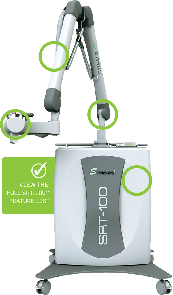
采用SRT-100浅层X线放射治疗仪,以瘢痕组织外扩0.5 cm为照射范围,每次照射剂量为4.5 Gy,1周照射1次,共4次。照射总剂量为18 Gy。治疗时创面周围贴放射线保护膜。
(四)局部药物注射治疗
将1 ml醋酸曲安奈德注射液(规格1 ml∶40 mg,浙江仙琚制药股份有限公司),0.6 ml氟尿嘧啶注射液(规格10 ml∶250 mg,上海旭东海普药业有限公司)和3.4 ml 2%盐酸利多卡因注射液制备成5 ml混合液 [ 7 , 8 ]。采用1 ml螺旋口注射器缓慢均匀注射至瘢痕疙瘩内,单次注射的终点反应为瘢痕疙瘩变白。1个月注射1次,共4次。
(五)2组治疗方式
观察组先进行环钻切除术,术后24 h内照射1次浅层X线,之后每周照射1次,共照射4次,放疗结束1周后予局部药物注射治疗,每月1次,共4次;对照组不接受环钻切除术,其他治疗与观察组相同。
三、观察指标
(一)瘢痕疙瘩总体积
于治疗前和疗程结束后12个月测量患者单个瘢痕疙瘩长、宽、厚度,计算单个瘢痕疙瘩体积,并求和计算每例患者的瘢痕疙瘩总体积,以cm 3为计量单位。
(二)局部注射药物总量
每次局部注射药物时记录实际注射量,4次治疗结束后计算每例患者局部注射药物总量,以ml为计量单位。
(三)VSS评分及有效性评价
于治疗前和治疗结束后3、6、12个月,分别对2组患者进行VSS评分,包括色泽(0~3分)、厚度(0~4分)、血管分布(0~3分)、柔韧性(0~5分)4个维度,总分0~15分,评分越高表示瘢痕越严重 [ 9 ]。疗程结束后12个月,依据VSS评分进行有效性评价,总分0分为治愈,1~5分为显效,6~10分为好转,11~15分为无效。显效率=(治愈+显效)例数/总例数×100%;有效率=(治愈+显效+好转)例数/总例数×100%。
(四)不良反应
记录整个研究过程中患者出现的所有不良反应。
四、统计学分析
采用SPSS 26.0软件进行数据分析,计量资料采用K-S法检验数据的正态性,符合正态分布的资料用x±s表示,2组间比较采用独立样本 t检验,同组间不同治疗时间点的比较采用重复测量数据的方差分析;计数资料用例(%)表示,组间比较采用 χ 2检验。P<0.05为差异有统计学意义。
结果
一、一般资料
......
除了消除手术后感染和复发的风险外,SRT-100还为患者和医生提供安全有效的治疗选择等多种优势,包括: